Studying Compound Mediated Effects on Primary Cultured Endothelial and Epithelial Cells
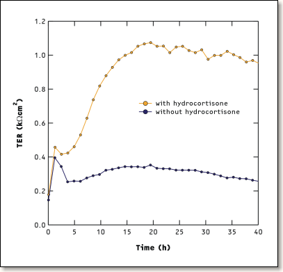
Time-resolved monitoring of the transepithelial electrical resistance (TER) of primary cultured endothelial cells derived from porcine brain microvessels, incubated in serum-free medium supplemented with hydrocortisone (orange curve) and without hydrocortisone (blue curve): the experimental data reveal that the TER of the confluent cell layer increases with time in the presence of hydrocortisone. This effect is attributed to a pronounced barrier strengthening of the cerebral endothelial cells.
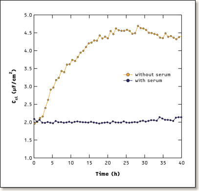
Time-resolved monitoring of the capacitance (Ccl) of primary cultured epithelial cells derived from porcine choroid plexus, incubated in serum-free medium (orange curve) and in serum-containing medium (blue curve): choroid plexus epithelial cells develop longer and more densely packed microvilli on their apical surface when incubated in a serum-free medium. This differentiation process leads to an increase of the capacitance of the confluent cell layer and can thus be followed non-invasively in situ.